RADIOLOGY CORNER |
https://doi.org/10.5005/jp-journals-10040-1225 |
Techniques for Ideal Intraoperative Radiography of Calcaneum
1,2Department of Orthopaedics, Postgraduate Institute of Medical Education and Research, Chandigarh, India
Corresponding Author: Shahnawaz Khan, Department of Orthopaedics, Postgraduate Institute of Medical Education and Research, Chandigarh, India, Phone: +91 9437536053, email: drskhan2018@gmail.com
ABSTRACT
Calcaneum is an important bone in the foot and ankle system. It is involved in the weight-bearing axis and also forms the arches of the foot. Any fracture of calcaneus needs to be treated. Operating on a fractured calcaneus is technically demanding and requires thorough knowledge of foot and ankle radiography. This helps in accurate reduction of the fracture and improves patient outcomes. To analyze an image of calcaneus, one must have knowledge about the anatomy and the angular measurements of the calcaneus. These structures are then visualized on radiographic images. Lateral view of foot, Harris axial view, Broden’s view, Saltzman’s view and the Captain’s view are some of the most frequently performed radiographs intraoperatively.
How to cite this article: Patel S, Khan S. Techniques for Ideal Intraoperative Radiography of Calcaneum. J Foot Ankle Surg (Asia Pacific) 2022;9(2):72-74.
Source of support: Nil
Conflict of interest: None
Keywords: Ankle, Calcaneus, Foot, Radiography, Radiographs, Trauma
INTRODUCTION
Calcaneus is an important bone of the foot lying along the weight-bearing axis, and also forms the posterior pillar of hindfoot serving as the solitary support to the arch of the foot. Any fracture of calcaneum if mismanaged can have devastating complications which in turn will affect the gait and locomotion of a human. Operating on fractures of calcaneus is both challenging and technically demanding, and the ideal goals of surgery are to restore the height of calcaneus, the correct alignment and anatomical reduction of the subtalar joint.
A thorough understanding of foot and ankle radiology is important to make an accurate diagnosis and avoid missed injuries that are common in foot and ankle. With the advent of CT scans, the various radiographic views of foot and ankle have become redundant. The teaching of the young budding surgeons is now more focused on studying CT scans. However, when it comes to operating a case, not all centers have the facility of getting intra-op CT scans. But the C-arm can readily provide radiographic images on OT table. The complex three-dimensional anatomy of the calcaneus can be interpreted by standard and special radiographs intraoperatively. Lateral view of foot, Harris axial view, Broden’s view and the Captain’s view are some of the most frequently performed radiographs intraoperatively. It is important for all Orthopaedic surgeons to know how to obtain radiographic views of foot and ankle intra-op.
To analyze an image of calcaneus, one must have knowledge about the anatomy and the angular measurements of the calcaneus.
Relevant Anatomy and Measurements
The calcaneus is a part of hindfoot. Broadly, it is composed of the anterior process, the calcaneal tuberosity posteriorly and the three facets: anterior facet, posterior facet, middle facet or the sustentaculum tali. It has numerous muscular and ligamentous attachments. It articulates with talus to form the subtalar joint and with cuboid bone anteriorly.1
On the lateral aspect, a bony elevation called peroneal tubercle is present which separates the tendons of peroneus longus from peroneus brevis. The sustentaculum tali forms the attachment of the tibio-calcaneal ligaments. The undersurface of sustentaculum tali is related to the tendon of flexor hallucis longus. Posteriorly on the plantar aspect, the calcaneal tubercle has two processes that serve as attachment of the plantar fascia. The posterior facet fractures show alterations in Bohler’s (Normal value = 25°-40°) and Gissane’s angle (Normal value = 130°-145). These angles are measured on lateral view radiographs as shown in Figures 1 and 2 .

Fig. 1: Bohler’s angle

Fig. 2: Gissane’s angle
There is a small tunnel located between the talus and the calcaneus on the lateral side. This tunnel is called the “Tarsal sinus.”2
Radiographic Views of Calcaneum
There are various radiographic views for identifying calcaneum fractures. Some of the most common radiographic views that are performed intraoperatively are:
Lateral View of Foot
A normal lateral view radiograph of foot helps in delineating the normal anatomy and shape of the bone. Fracture morphology can also be made out (tongue type or joint depression type) in this view of radiograph. In this view, one should also look for Bohler’s angle and Gissane angle. Any alteration in angle suggests posterior facet displacement or dislocation.
Procedure: In lateral position (commonest position for surgery), the C-arm is brought perpendicular to the foot and the X-ray beam is fired (Fig. 3).
Figs 3A and B: Correct technique of obtaining lateral view and C-arm shot of lateral view
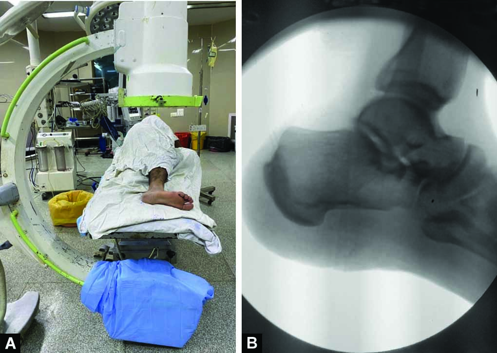
Harris Axial View
Originally described by Harris and Beath to identify talocalcaneal coalition in a rigid flat foot, this view helps us to assess the axial alignment of the calcaneus and identify medial or lateral shift of fracture fragments.3 It also helps to assess the integrity of the subtalar joint and the heel width.
Procedure
-
The patient is usually operated in a lateral position. The operative limb is externally rotated at the hip so that the toes point upwards to the sky.
-
The foot is dorsiflexed until the plantar surface is perpendicular to the image receptor and then the X-ray beam is fired (The correct technique and intraoperative image is shown in Figure 4).

Figs 4A and B: Correct technique of obtaining Harris axial view and C-arm shot of Harris axial view
Broden’s View
This view helps us to check the reduction of the posterior facet.1
Procedure
-
The patient is in a lateral position. The operative limb is then externally rotated at the hip such that the toes point upwards to the sky.
-
The X-ray beam is directed towards the foot at an angle of 20-40°.
-
The foot is internally rotated at 10, 20, 30, and 40° and the X-ray beam is fired to visualize the posterior facet. The correct technique and intraoperative image is shown in Figure 5.

Figs 5A and B: Correct technique of obtaining Broden’s view and C-arm shot of Broden’s view
Captain’s View
This view is also used to evaluate the axial alignment of the calcaneum.4,5 This method demonstrated more accurate measurement of increasing heel varus compared with the Harris view. The correct technique and intraoperative image is shown in Figure 6.

Figs 6A and B: Correct technique of obtaining Captain’s view and C-arm shot of Captain’s view
Procedure
-
The patient is placed in a lateral position.
-
The operative lower limb is externally rotated at the hip joint such that the toes face the sky.
-
The ankle is placed in neutral rotation and then maximally plantarflexed till the foot is perpendicular to the X-ray beam.
-
The X-ray beam is then fired centring over the talus.
Saltzman’s View
This view helps to assess the hindfoot malalignment after performing calcaneal osteotomies.6 The correct technique and intraoperative image is shown in Figure 7.

Figs 7A and B: Correct technique of obtaining Saltzman’s view and C-arm shot of Saltzman’s view
Procedure
-
The patient is placed in a lateral position.
-
The C-arm is aligned in such a way that the collimator is angled at an angle of 20° from horizontal.
-
The beam is centered over the distal tibia and ankle joint.
-
The long axis of the tibia and calcaneus give the malalignment angle.
REFERENCES
1. Brian PL, Mahraj RPM. Imaging of the calcaneus. Foot Ankle Clin 2005;10(3):443-461, vi. DOI: 10.1016/j.fcl.2005.04.002
2. Yamaguchi R, Nimura A, Amaha K, et al. Anatomy of the tarsal canal and sinus in relation to the subtalar joint capsule. Foot Ankle Int 2018;39(11):1360-1369. DOI: 10.1177/1071100718788038
3. Harris RI, Beath T. Etiology of peroneal spastic flat foot. J Bone Joint Surg Br 1948;30B(4):624-634. DOI: 10.1302/0301-620X.30B4.624
4. Kwon JY, Moura B, Gonzalez T, et al. Anterior-Posterior (AP) Calcaneal Profile View: A Novel Radiographic Image to Assess Varus Malalignment. Foot Ankle Int 2020;41(10):1249-1255. DOI: 10.1177/1071100720937297
5. Velasco B, Moura B, Kwon J. The captain’s view: a novel radiographic view to assess axial alignment of the calcaneus. Foot Ankle Orthop 2019;4(4):2473011419S00426. DOI: 10.1177/2473011419S00426
6. Saltzman CL, El-Khoury GY. The Hindfoot Alignment View. Foot Ankle Int 1995;16(9):572-576. DOI: 10.1177/107110079501600911
________________________
© The Author(s). 2022 Open Access This article is distributed under the terms of the Creative Commons Attribution 4.0 International License (https://creativecommons.org/licenses/by-nc/4.0/), which permits unrestricted use, distribution, and non-commercial reproduction in any medium, provided you give appropriate credit to the original author(s) and the source, provide a link to the Creative Commons license, and indicate if changes were made. The Creative Commons Public Domain Dedication waiver (http://creativecommons.org/publicdomain/zero/1.0/) applies to the data made available in this article, unless otherwise stated.